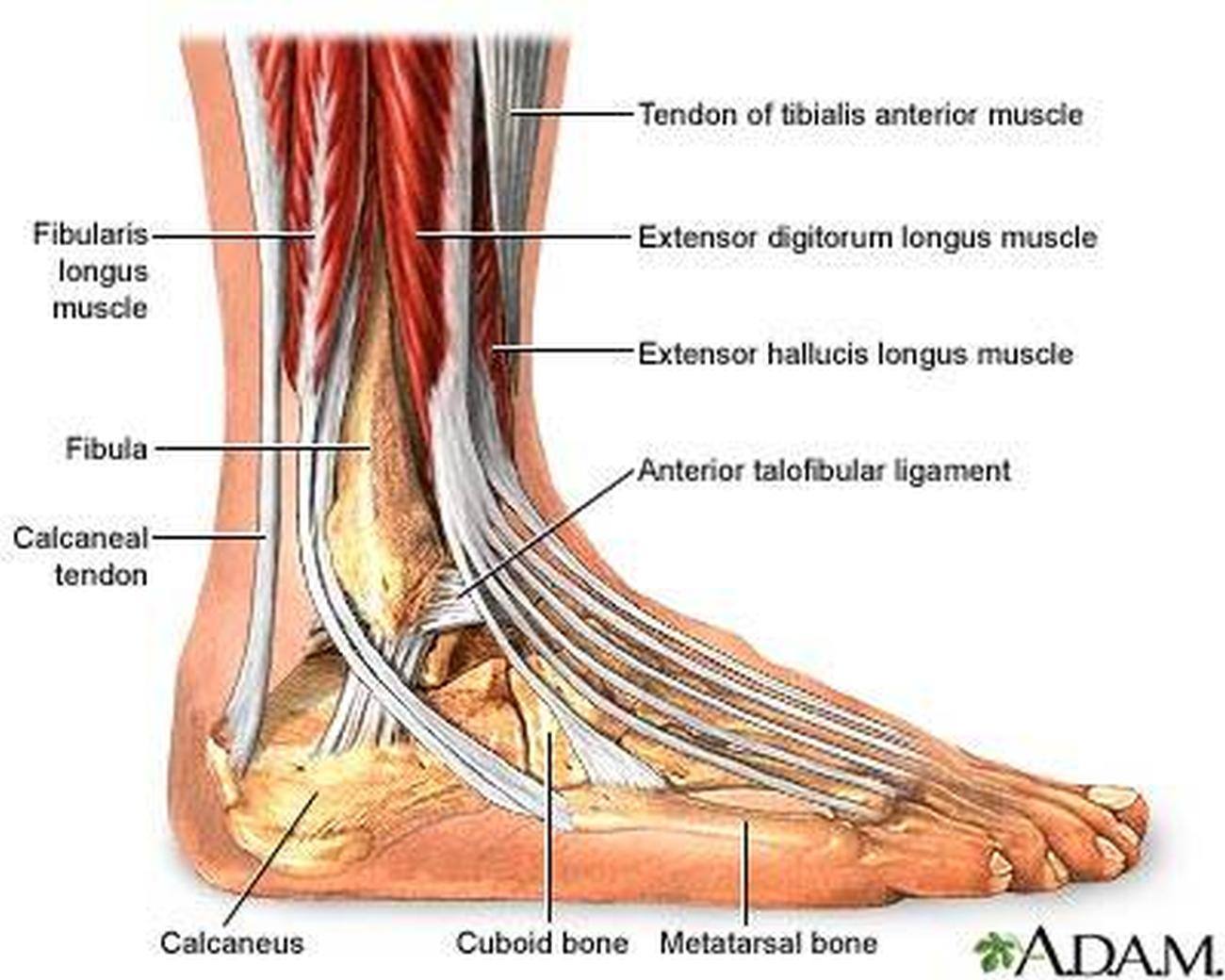
The Ankle Joint Biology Diagrams The anterior inferior tibial fibular ligament is commonly known as the "high ankle sprain ligament". It is positioned on the lower front of the ankle and helps keep the tibia and fibula together. Injuries to this ligament occur when the foot is stuck on the ground and rotated inwardly. The anterior inferior tibiofibular ligament (Figure 4) is positioned on the anterolateral aspect of the ankle joint and serves to helps keep the tibia and fibula together. Injuries to this ligament, so called high ankle sprains, occur when the foot is stuck on the ground while the leg rotates inwards. The Interosseous Membrane
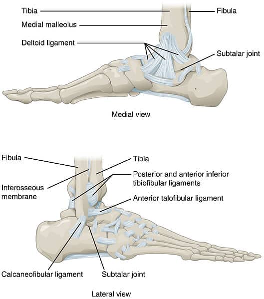
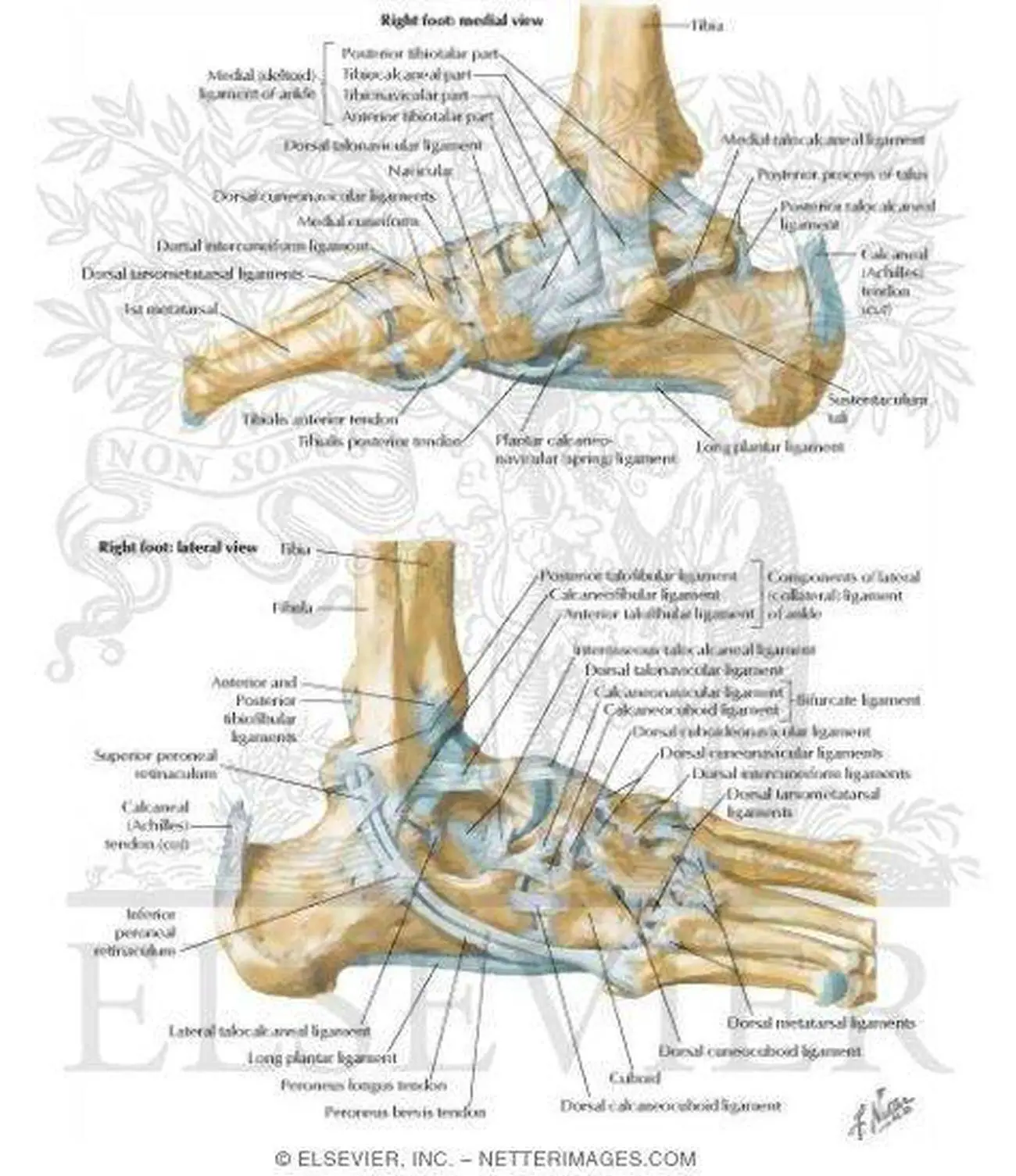
Ligaments [edit | edit source]. Acute and chronic ankle pain are often associated with either ligament injury or ligament laxity. The ligaments of the ankle consist of the medial ligaments (the deltoid ligament), the lateral ligaments, and the ligaments connecting the distal epiphyses of the tibia and fibula called the tibiofibular syndesmosis.
Ankle Ligaments: What They Are, Anatomy & Function Biology Diagrams
The interosseous ligament, which rests between the tibia and fibula and runs the entire length of the tibia and fibula, from the ankle to the knee; The various ligaments that surround the ankle together help form part of the joint capsule, a fluid-filled sac that surrounds and lubricates articulating joints. To prevent foot ligament injuries, it's important to strengthen the muscles that support and stabilize your ankle. Foot ligament strains often happen when you roll or twist an unstable, weak ankle. Other prevention measures include: Slowly increasing the duration and intensity of activity over time;
Ankle anatomy. The ankle joint, also known as the talocrural joint, allows dorsiflexion and plantar flexion of the foot. It is made up of three joints: upper ankle joint (tibiotarsal), talocalcaneonavicular, and subtalar joints.The last two together are called the lower ankle joint. The upper ankle joint is formed by the inferior surfaces of tibia and fibula, and the superior surface of talus.

Ankle Anatomy: Muscles and Ligaments Biology Diagrams
The foot and ankle form a complex system which consists of 28 bones, 33 joints, 112 ligaments, controlled by 13 extrinsic and 21 intrinsic muscles.. The foot is subdivided into the rearfoot, midfoot, and forefoot. It functions as a rigid structure for weight bearing and it can also function as a flexible structure to conform to uneven terrain. A thick ligament, called the deltoid ligament, supports the medial ankle (the side closest to your other ankle). Ligaments also support the lower end of the leg where it forms a hinge for the ankle. This series of ligaments supports the ankle syndesmosis, the part of the ankle where the bottom end of the fibula meets the tibia. Three main